Infrared Spectroscopy of cyanobacterial cells
measured on station13.3
at the Daresbury Laboratory
more details about Infrared Spectroscopy
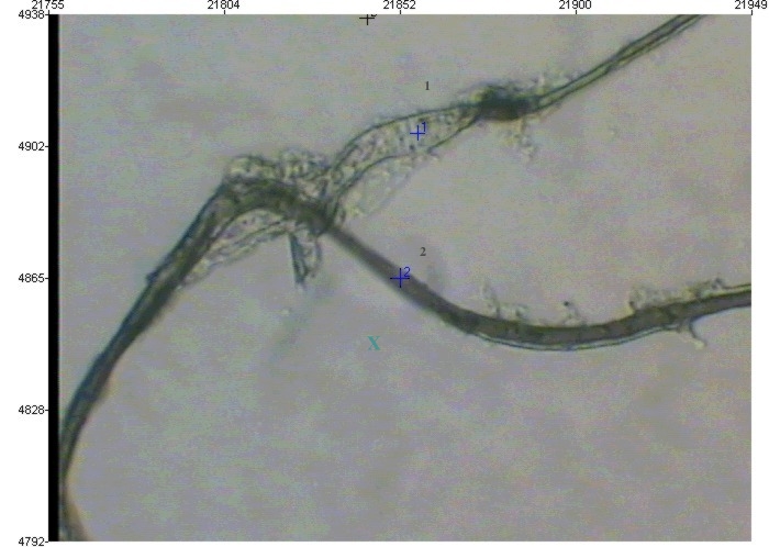
Fig. 1. Infrared image of Calothrix sp. that was silicified in situ at Krisuvik springs, Iceland. Silicification experiment was carried out for 4 days. Point 1 is on a bacterial sheath dislocated from the mother cell and corresponds to the IR spectra # 2 in (Figure below). Point 2 shows the site where the bacterium lost most of its sheath and corresponds to spectra # 5 in (Figure below)]; green cross represnets the background that was substracted in all the traces below.
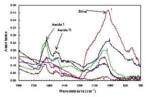
Fig. 2. Fourier transformed IR spectra of various Calothrix and standards. [starting from the top scan: red = silica standard; blue = Calothrix sheath silicified for 4 days at Krisuvik springs; green = Calothrix sheath silicified in the lab. for 6 days; purple = Calothrix with sheath - Krisuvik 4 days; black = Calothrix with no sheath - Krisuvik 4 days; brown = bacterial control, no silica.]